Electron micrograph of animal cell information
Home » Trend » Electron micrograph of animal cell informationYour Electron micrograph of animal cell images are available. Electron micrograph of animal cell are a topic that is being searched for and liked by netizens now. You can Download the Electron micrograph of animal cell files here. Find and Download all royalty-free images.
If you’re searching for electron micrograph of animal cell images information related to the electron micrograph of animal cell interest, you have come to the ideal blog. Our site frequently provides you with suggestions for seeking the maximum quality video and image content, please kindly surf and find more informative video content and graphics that fit your interests.
Electron Micrograph Of Animal Cell. The following sizes are ranges only. 1.2 is a diagram of an electron micrograph of an animal cell. 1.1 is a diagram of an electron micrograph of a plant cell. They are spherical vesicles which contain hydrolytic enzymes that can break down virtually all kinds of biomolecules (waste.
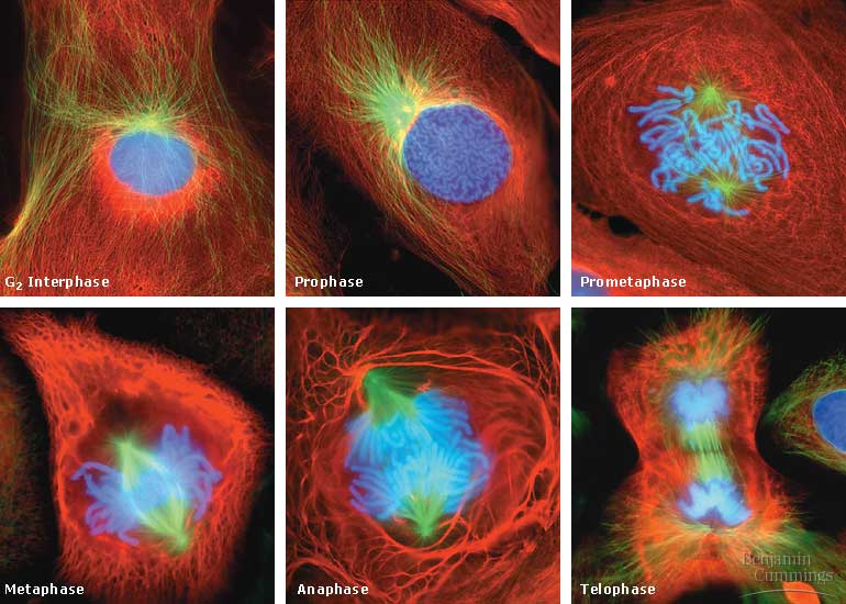
 The Cell Cycle From bio.utexas.edu
They have many mitochondria to supply the atp needed for these processes. The dark zone (centre right) in the nucleus is the nucleolus. Chromosomes figure 23 a cell in mitosis (telophase): Transmission electron micrograph (tem) of lysosomes. But at the same time it is interpretive. The electron micrograph displayed below illustrates many of the plant cell characteristics discussed the cell wall, large central vacuole and chloroplasts are clearly visible also visible is the clearly defined nucleus containing chromatin nucleus chromatin the vacuole in this mature plant cell from a leaf is large, and occupies about 80% of
(1) nucleus was discovered by brown (1831).
Chromosomes figure 23 a cell in mitosis (telophase): They have many mitochondria to supply the atp needed for these processes. Year 11 bio key points cell organelles and their function animal cell cell organelles eukaryotic cell. Animal and plant cells have certain. The electron micrograph displayed below illustrates many of the plant cell characteristics discussed the cell wall, large central vacuole and chloroplasts are clearly visible also visible is the clearly defined nucleus containing chromatin nucleus chromatin the vacuole in this mature plant cell from a leaf is large, and occupies about 80% of Typical animal cell pinocytotic vesicle lysosome golgi vesicles golgi vesicles rough er (endoplasmic reticulum) smooth er (no ribosomes) cell (plasma) membrane mitochondrion golgi apparatus nucleolus nucleus centrioles (2) each composed of 9.
 Source: sciencelearn.org.nz
An electron micrograph of a mouse liver cell dna learning center electrons cell learning centers. They have many mitochondria to supply the atp needed for these processes. Diagram of animal cell under electron microscope. Typical animal cell pinocytotic vesicle lysosome golgi vesicles golgi vesicles rough er (endoplasmic reticulum) smooth er (no ribosomes) cell (plasma) membrane mitochondrion golgi apparatus nucleolus nucleus centrioles (2) each composed of 9. Year 11 bio key points cell organelles and their function animal cell cell organelles eukaryotic cell.
 Source: bio.utexas.edu
Taking up most of the cell is the nucleus, where genes are stored in the form of chromosomes. The dark area in the nucleus is the nucleolus. This is the most active part of the nucleus, and contains unravelled chromosomes involved in making. The subcellular organelles of the host cell [mitochondria and plasts (a)] are present at the periphery of the cell adjacent to the host cell wall (b). Animal cell electron micrograph labeling.
 Source: turbosquid.com
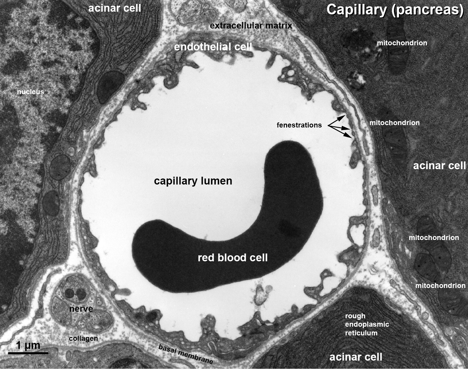
Figure 21 epithelial cells often display extensive basal plasma membrane infoldings as observed in this electron micrograph: The dark area in the nucleus is the nucleolus. Animal cell electron micrograph labeling. Diagram of animal cell under electron microscope. Animal and plant cells have certain.
 Source: embryology.med.unsw.edu.au
List ten structures you could find in an electron micrograph of an animal cell which would be absent from the cell of a bacterium. Learn vocabulary, terms, and more with flashcards, games, and other study tools. Transmission electron micrograph (16,000x enlargement) through an infected root cell of a soybean plant. Animal and plant cells have certain. But at the same time it is interpretive.
 Source: sciencephoto.com
Cell structure light and electron microscopes allow us to see inside cells. But at the same time it is interpretive. List ten structures you could find in an electron micrograph of an animal cell which would be absent from the cell of a bacterium. They will have all the organelles of an animal cell but will have many ribosomes and rough er to create the enzymes which are proteins and transport them outside the cell. Seen under the electron microscope (not to scale) one function of organelle cell type(s) in which organelle is located mitochondrion animal and plant assemble microtubules to produce the mitotic spindle rough endoplasmic reticulum protein synthesis golgi apparatus animal and plant photosynthesis plant only [8] [total:
 Source: embryology.med.unsw.edu.au
This is an electron micrograph of nucleus. I pinimg com 474x d7 30 97 d730978ce46b9f67e308. They are spherical vesicles which contain hydrolytic enzymes that can break down virtually all kinds of biomolecules (waste. The actual diameter of the animal cell is 45 um.) the observed diameter of the animal cell in the electron micrograph is 1.8 cm. Pin by nia on education plant cell electron microscope cell.
This site is an open community for users to submit their favorite wallpapers on the internet, all images or pictures in this website are for personal wallpaper use only, it is stricly prohibited to use this wallpaper for commercial purposes, if you are the author and find this image is shared without your permission, please kindly raise a DMCA report to Us.
If you find this site convienient, please support us by sharing this posts to your preference social media accounts like Facebook, Instagram and so on or you can also save this blog page with the title electron micrograph of animal cell by using Ctrl + D for devices a laptop with a Windows operating system or Command + D for laptops with an Apple operating system. If you use a smartphone, you can also use the drawer menu of the browser you are using. Whether it’s a Windows, Mac, iOS or Android operating system, you will still be able to bookmark this website.
Category
Related By Category
- Anime like cowboy bebop information
- Best anime gifs information
- Do animals cry information
- Arc animal rescue information
- Anime thriller genre information
- Dyson v7 animal black friday information
- Copyright free cartoon animal images information
- Fantastic four the animated series episodes information
- Dyson v11 animal black friday 2019 information
- Coniferous forest animals information